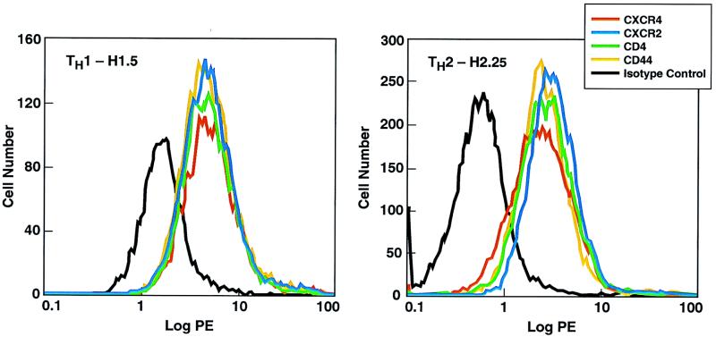
FIG. 2.
Flow cytometric analysis of a human TH1 clone and a human TH2 clone for cell surface expression of chemokine receptors. A total of 106 cells of each of the T-cell clones H1.5 and H2.25 were suspended in PBS containing 1% heat-inactivated human AB serum and 0.5% sodium azide and stained with phycoerythrin (PE)-labeled anti-CXCR2, -CXCR4, -CD4, or -CD44 or an isotype control labeled immunoglobulin G antibody. After staining, the cells were extensively washed and then fixed with 1% paraformaldehyde. The clones were analyzed on a FACStar Plus flow cytometer.

