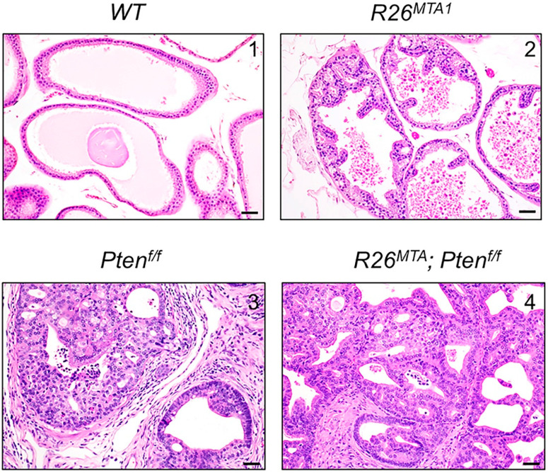
Figure 1.
Representative photomicrographs of prostates from four prostate-specific genetic groups: (1) R26MTA1; Pb-Cre-negative (WT), (2) R26MTA1, (3) Ptenf/f, and (4) R26MTA1; Ptenf/f were evaluated using H&E staining (scale bar, 100 µm). The prostate in WT mice is unremarkable (upper left). In R26MTA1 mice, there is an occasional hyperplasia of the prostatic epithelium without nuclear or cellular atypia (upper right). In Ptenf/f and R26MTA1; Ptenf/f mice, both PIN and prostatic adenocarcinoma are observed. The images (lower left and right) show well to moderately differentiated invasive prostatic adenocarcinoma with desmoplasia and inflammatory infiltrates.

