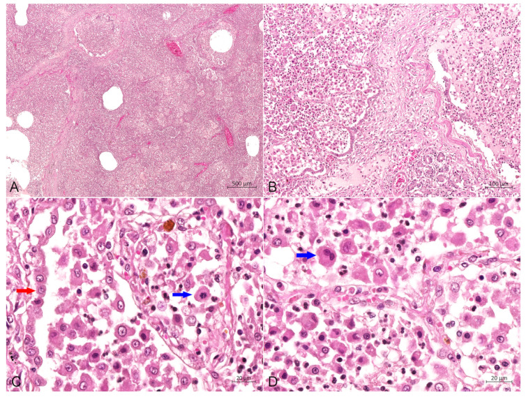
Figure 4.
Hematoxylin-eosin stain of the lung. (A) The lobular, bronchiolar and alveolar structures of the lung disappeared and were filled with a large number of PHCs, bar = 500 μm. (B) Proliferation of numerous histiocytic cells in the alveolar cavity (left) and bronchioles (right), with severe fibrosis in the interstitium, bar = 100 μm. (C) The alveolar cavity was repleted of PHCs, with occasional mitotic figures (blue arrow), and type II alveolar epithelial cells (red arrow) showed prominent hyperplasia, bar = 20 μm. (D) A large number of PHCs in the alveolar cavity with occasional binucleated cells (blue arrow), bar = 20 μm.

