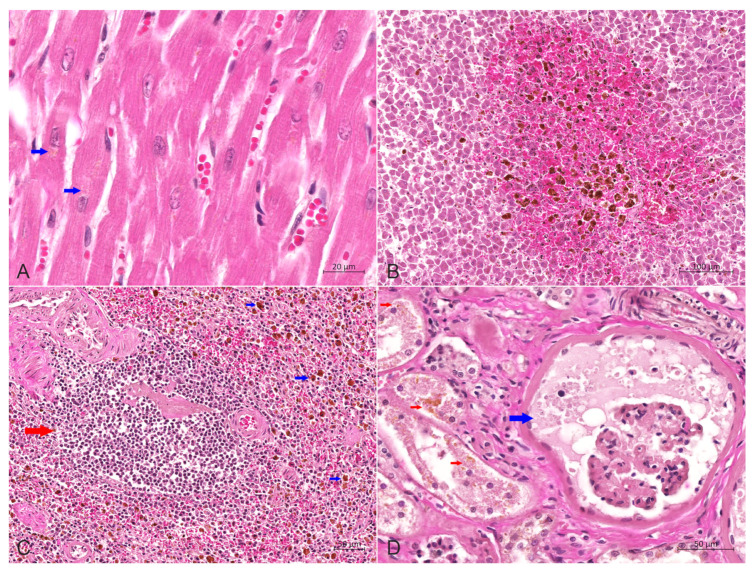
Figure 5.
Hematoxylin-eosin stain of heart, liver, spleen and kidneys. (A) Slight congestion and dilation of the small blood vessels in the myocardial interstitium were observed, with a small amount of brownish yellow lipofuscin particles (red arrow) in the cytoplasm of myocardial cells, bar = 20 μm. (B) The central area of the liver lobules was significantly congested, and the activation and proliferation of Kupffer cells containing hemosiderin were increased, bar = 100 μm. (C) The number of lymphocytes in the spleen was reduced considerably, and the volume of white pulp (red arrow) was decreased. Also, macrophages containing hemosiderin (blue arrow) were dispersed in the red pulp, bar = 50 μm. (D) Wide renal fibrosis was seen, with the extensive proliferation of connective tissue around some glomeruli, dilation of renal sacs filled with protein-rich urine (blue arrow), degeneration of renal tubular epithelial cells, and noticeable lipofuscin particles (red arrow) in the cytoplasm, bar = 50 μm.

