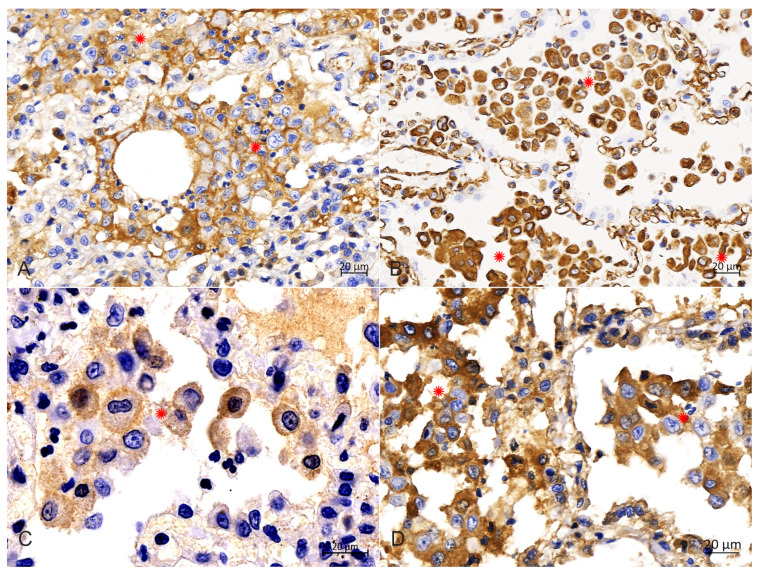
Figure 6.
Immunohistochemical analysis in the cytoplasm of PHCs. (A) Immunohistochemical staining of the CD1a was strongly positive in the membrane of PHCs (around asterisks), bar = 20 μm. (B) Immunohistochemical staining of vimentin was strongly positive in the cytoplasm of PHCs (around asterisks), bar = 20 μm. (C) Immunohistochemical staining of S100 was strongly positive in the intramembrane of PHCs (around asterisks), bar = 20 μm. (D) Immunohistochemical staining of E-cadherin was strongly positive in the cytoplasm of PHCs (around asterisks), bar = 20 μm.

