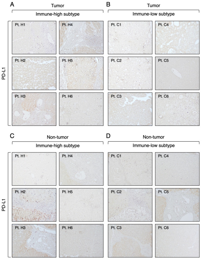
Figure 9.
Expression of PD-L1 molecule in the tumor and nontumorous tissue of immune-high and immune-low subtypes. The images illustrate the immunostaining with monoclonal antibody against PD-L1 in paraffin liver sections taken at the time of liver transplantation from the center of the tumor (A,B) and the surrounding nontumorous tissue (C,D). The left two columns show the immune-high subtype, and the right two columns show the immune-low subtype (100× magnification).

