Figure 5.
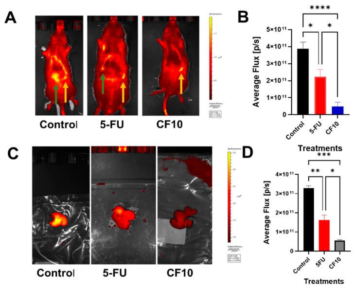
Imaging CRLM progression in WAG/Rij rats after 4 weeks of the indicated treatment, showing that CF10 was found to be effective in a rat CRLM model. Liver metastases were formed by injecting CC531 rat CRLM cells into the left hepatic lobe. Liver tumor masses were detected using a fluorescent (Cy5.5-labeled) RGD peptide and were similar in all rats at baseline (2 weeks after receiving an injection). Rats (n = 5 per group) were then treated for 4 weeks (1×/week via i.v. dose) with vehicle, 5-FU (50 mg/kg), or CF10 (identical to 50 mg/kg 5-FU dose based on UV A260). (A) The fluorescence imaging of rats 4 weeks after receiving treatment. Green arrows point to liver tumor signal, which is quantified in (B). Yellow arrows point to spleen autofluorescence. (C) Rats were then euthanized, and their livers were extracted and imaged ex vivo with Cy5.5 signal quantified in (D). Rats treated with CF10 displayed significantly reduced signals from CRLMs relative to both 5-FU and vehicle. * p < 0.01; ** p < 0.011; *** p < 0.015; **** p < 0.002.

