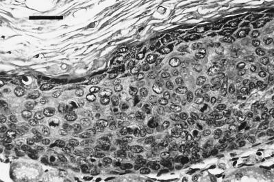
FIG. 3.
First isolation. Histology (hematoxylin-eosin stain) of one of the renal grafts from the third passage experiment shows prominent signs of intraepithelial neoplasia with basaloid proliferation, nuclear pleomorphism, dyskeratosis, and multiple aberrant mitoses throughout the stratum spinosum (bar, 100 μm).

