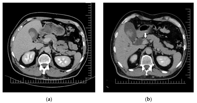
Figure 3.
Biliary duct metastases: (a) Polypoid gallbladder metastasis on initial CT. (b) Dilation of cholecyst (containing multiple enlarged polypoid lesions) and extrahepatic bile ducts secondary to distal common bile duct obstruction though iodophilic melanoma metastasis (arrow). Scale bar: 12.5 mm.

