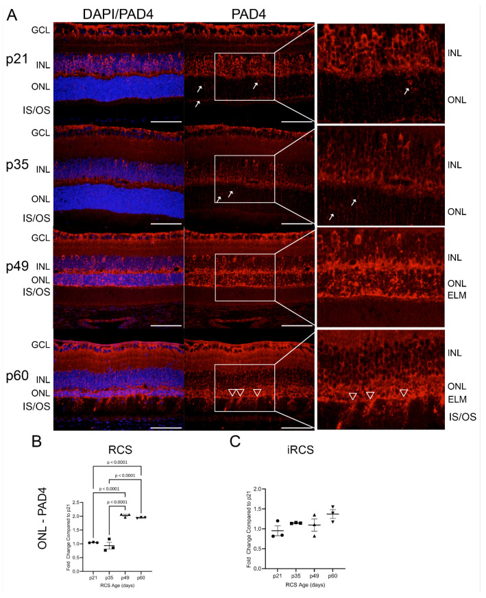
Figure 8.
Representative immunofluorescence staining of PAD4 in RCS retinas from p21 to p60; 40× images of DAPI (blue) and PAD4 (red) (A). Early at p21, PAD4 is retained in the ganglion cell layer (GCL) and inner nuclear layer (INL) but focal staining in the ONL is observed at p21 and p35 (white arrows); by p49 strong expression is seen in outer nuclear layer (ONL) up to external limiting membrane (ELM), and at p60 there are regions of the ELM where PAD4 extends into the inner and outer segments (IS/OS, white triangles). ImageJ analysis show significant ONL increase in PAD4 expression for RCS rats (B), but not for iRCS rats (C). Data represented as mean ± SEM, one-way ANOVA with Tukey’s correction, n = 3 each age group p21 (circle), p35 (squares), p49 (triangles), and p60 (inverted triangles) (B,C). Scale bar = 50 µm.

