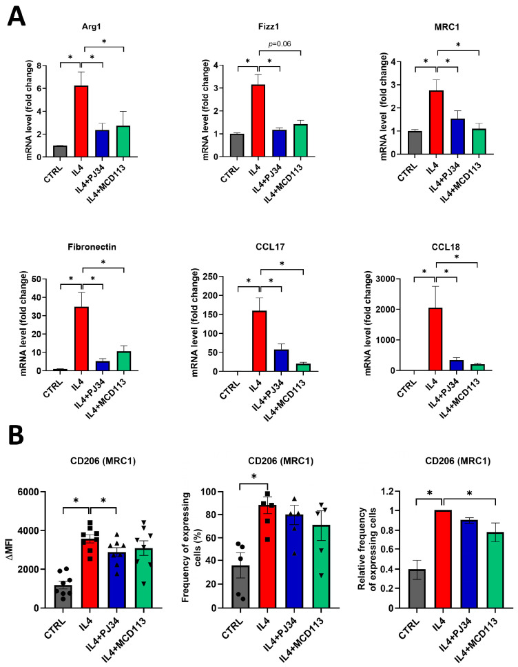
Figure 3.
The PARP inhibitor PJ34 and the PARP14 inhibitor MCD113 reduced the expression of M2 markers in human macrophages. Macrophages were differentiated from human monocytes with 50 ng/mL M-CSF for 5 days. On day 5, they were pre-treated with either 20 μM PJ34 or 50 μM MCD113 for 1 h and then stimulated with 100 ng/mL IL-4 for 24 h to polarize them into M2 macrophages. On day 6, cells were collected for RNA extraction, and M2 markers were measured with RT-qPCR. Histograms show the mean of at least 3 independent experiments (±SEM). Statistical evaluation was performed with the Kruskal–Wallis test and Dunn’s post-hoc test (* p < 0.05) (A). On day 6, cells were also collected and labelled with APC-conjugated anti-human mouse CD206 monoclonal antibody. Fluorescence intensities and the ratio of CD206-expressing cells were measured by flow cytometry. The mean value of MFIs (median fluorescence intensities) of the cell surface molecule CD206 was calculated from 8 independent experiments (±SEM). The mean value of frequency of CD206+ cells was calculated from 5 independent experiments (±SEM). The relative frequency of CD206-expressing cells was determined as follows. For each donor, the frequency of CD206+ cells differentiated in the presence of IL-4 was considered to be 1, and the frequency of untreated cells and cells treated with IL-4 + PARP inhibitors was compared to this. Data were analyzed by one-way ANOVA followed by Bonferroni’s post-hoc test (left panel) or by the Kruskal–Wallis test and Dunn’s post-hoc test (middle and right panels) (* p < 0.05) (B).

