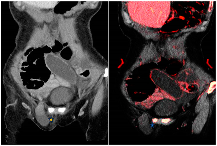
Figure 1.
From left to right—Contrast enhanced conventional 120 kVp and iodine overlay map (IOM) coronal images of the abdomen. Closed-loop small bowel obstruction secondary to a right inguinal hernia (*). The small bowel loop demonstrates no mural enhancement; more conspicuous on the IOM (*) which is highly suggestive of an incarcerated hernia.

