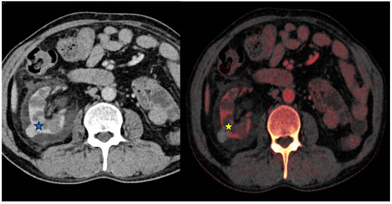
Figure 3.
From left to right—Contrast-enhanced (portal venous) and Iodine overlay map (IOM) axial images of the abdomen. Right exophytic hyperattenuating lesion (*) is further characterized as lacking internal enhancement on the IOM (*) image and is therefore likely to represent a benign hyperdense cyst rather than an enhancing lesion.

