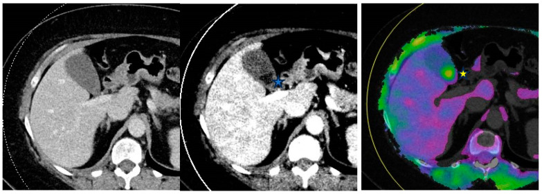
Figure 4.
From left to right—Conventional mixed 120 kVp equivalent, 40 keV virtual monoenergetic and gallstone analysis application axial images through the abdomen. Gallstones are not visible on the conventional image as they isoattenuate to the surrounding bile. However, they are visible on the DECT images and appear hypodense (filling defect) on the virtual monoenergetic image (*) and hyperdense (*) on the gallstone analysis application.

