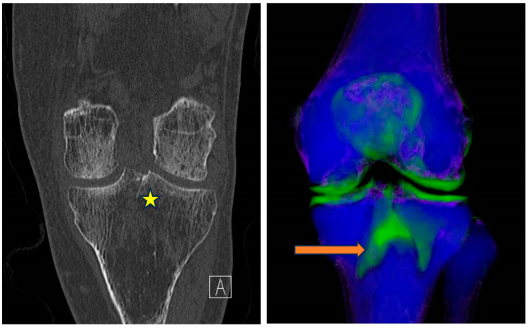
Figure 6.
From left to right—Conventional mixed 120 kVp equivalent and 3D bone marrow edema overlay coronal images of the right knee. Subtle intercondylar tibial fracture extending into the tibial condyle (*) is confidently diagnosed when appreciating the associated BME on the overlay image (orange arrow).

