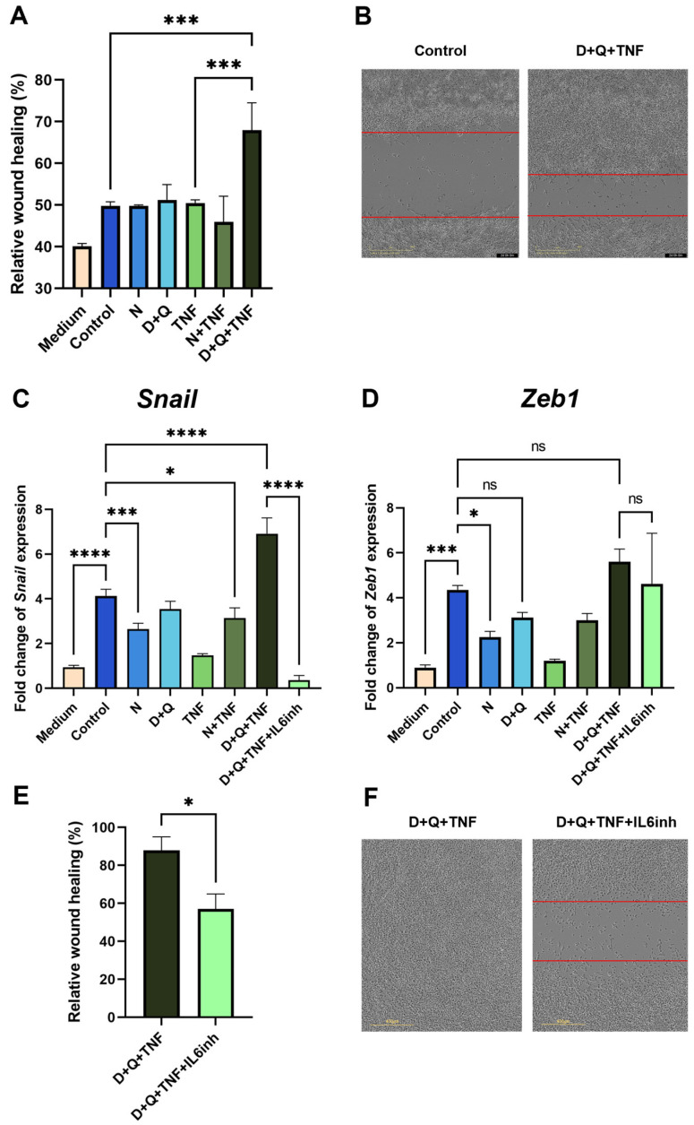
Figure 5.
CAFs treated with the D + Q combination promoted the expression of EMT markers and enhanced the migration of MC38 cancer cells. (A) Analysis of cell migration activity in the scratch wound healing assay quantified using the IncuCyte live-cell imaging system. Mean migration/invasion values ± SEM (n = 3). (B) Representative images of MC38 cells in the scratch wound healing assay at 0 and 48 h in areas with the addition of CM from untreated CAFs and CAFs treated with D + Q + TNF. (C) Expression of Snail and (D) Zeb1 in CAFs was analyzed by qPCR. Charts represent relative expression levels as mean values ± SEM normalized by Gapdh. Data from one of three independent experiments are shown. (E) Analysis of cell migration activity in the scratch wound healing assay. Mean migration/invasion values ± SEM (n = 3). (F) Representative images of MC38 cells in the scratch wound healing assay at 72 h in areas with the addition of CM from CAFs treated with D + Q + TNF with or without anti-IL-6 antibodies (IL-6inh). In all images scale bar 400 μm. Data were analyzed using one-way ANOVA with Tukey’s multiple comparisons test; * p < 0.05, *** p < 0.001, **** p < 0.0001, ns—not significant.

