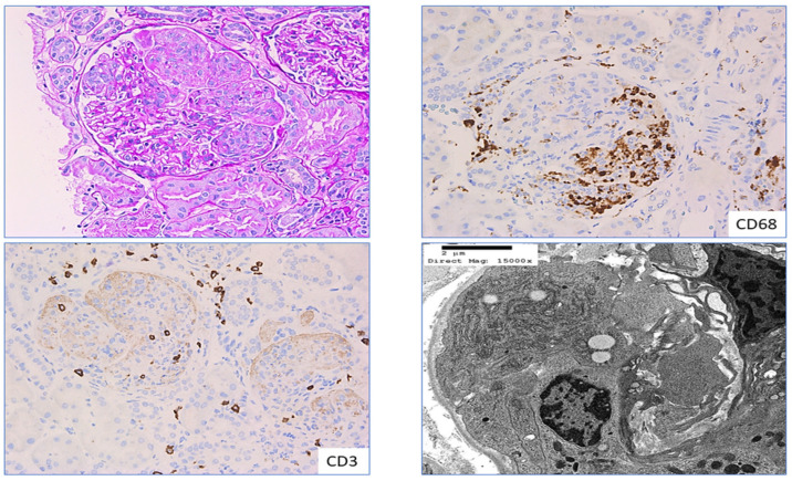
Figure 5.
CD3 and CD68 in pauci-immune crescentic glomerulonephritis: Section from a patient with pauci-immune crescentic glomerulonephritis shows cellular crescent in a glomerulus with collapsed tuft exhibiting focal fibrinoid necrosis (left top pane); left bottom pane and right top pane highlights infiltration of CD3-positive T lymphocytes and CD68-positive macrophages in the same glomerulus, ×200; right bottom pane highlights ultrastructure of macrophage infiltration in the capillary loop of the glomerulus from the same patient.

