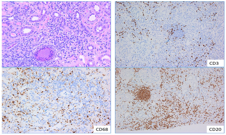
Figure 6.
Monocytes and macrophages in tubulointerstitial nephritis.—Sections from a patient with drug-induced tubulo-interstitial nephritis highlights the density of lympho-plasmacytic and histiocytic infiltration in the renal interstitium, ×200 (left top pane). Images in the left bottom and right pane highlights immunoperoxidase stains with CD68, CD3 and CD20 demonstrating the distribution of macrophages, T and B lymphocytes, respectively in the interstitial infiltrate, ×100.

