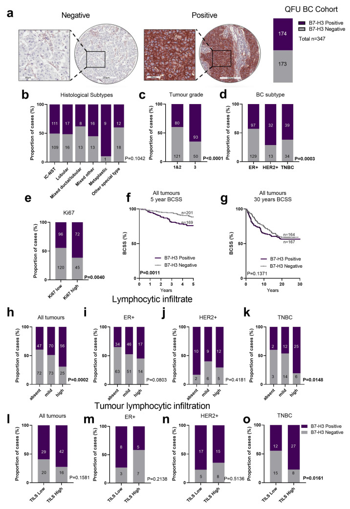
Figure 2.
B7-H3 expression in breast tumours in the QFU cohort. (a) Representative immunohistochemistry images of breast tumour cores scored as negative (left image) or positive (right image) for B7-H3 expression with 50% of the breast tumours in the QFU cohort stained positive for B7-H3 (n = 173 of 343). (b) Chi-square analysis of the proportion of breast cancer histological subtypes with negative or positive B7-H3 expression. (c) Chi-square analysis of the proportion of breast cancer cases with positive and negative B7-H3 expression compared across tumour grades. (d) Chi-square analysis of the proportion of breast cancer subtypes (ER+, HER2+, and TNBC) with negative and positive B7-H3 expression. (e) Chi-square analysis of B7-H3 expression in Ki67-low and Ki-67-high cases across all breast cancer subtypes. (f,g) Kaplan–Meier (KM) analysis of B7-H3 expression and 5-year (f) and 30-year BCSS (g). (h) Chi-square analysis of lymphocytic infiltration in all tumour subtypes, and in ER+ only (i), HER2+ only (j), and TNBC only (k). (l) Chi-square analysis of tumour infiltrating lymphocytes (TILS) in all tumour subtypes, and: ER+ only (m), HER2+ only (n), and TNBC only (o). BC, breast cancer; BCSS, breast cancer specific survival; ER+, oestrogen receptor; HER2+, human epidermal growth factor receptor 2; TILs, tumour infiltrating lymphocytes; TNBC, triple negative breast cancer. Scale bar: core = 200 µm; inset 60 µm.

