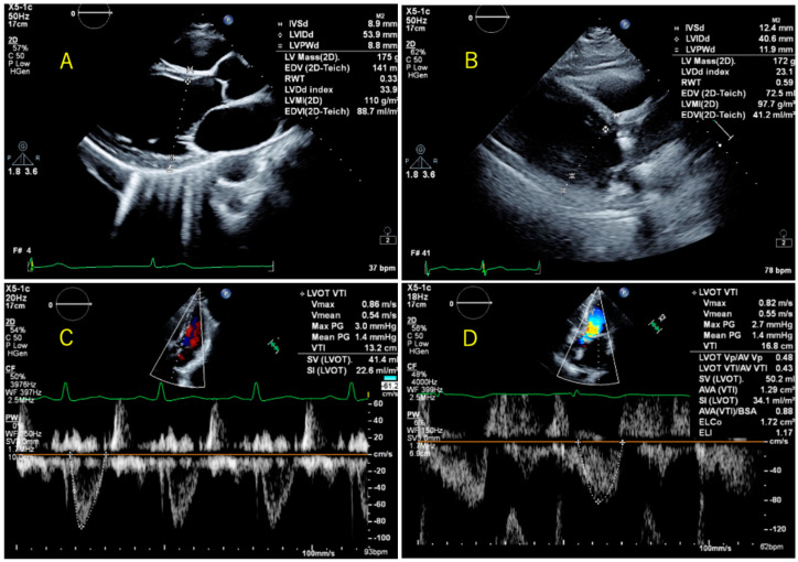
Figure 1.
Representative cases where fully automatic measurement was possible (A,C), and cases where correction was necessary (B,D). (A) had good image quality and did not need to be corrected after automatic measurement. (B) was of poor image quality, and the measurement position did not capture the boundaries of the left ventricle, so a correction was made. (C) No correction was made after automatic measurement. (D) Corrections were made because the boundaries of the pulsed Doppler waveform were not captured.

