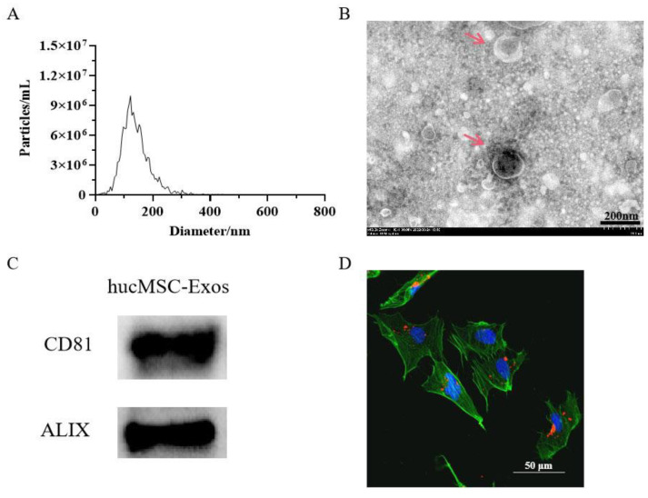
Figure 1.
Characterization of exosomes derived from human umbilical cord mesenchymal stem cells (HucMSC-Exos). (A) Nanoparticle tracking analysis (NTA) of HucMSC-Exos. The mean diameter of HucMSC-Exos was 141 nm. (B) Transmission electron microscopic (TEM) analysis of exosomes; The red arrows point to the HucMSC-Exos; scale bars are 200 nm. (C) Immunoblotting for CD81 and ALIX in exosomes. (D) Verification of the uptake of exosomes in skin cells. HucMSC-Exos were stained with PKH26® (red) and incubated with human skin fibroblasts (HSFs) for 12 h. Before analysis, cells were counterstained with phalloidin (green) and nuclei were stained with 4,6-diamidino-2-phenylindole (DAPI) (blue) for counterstaining; scale bars are 50 µm.

