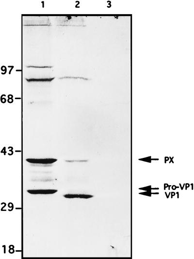
FIG. 8.
HAV VP1 immunoblot analysis following transfection with HAV cDNA. Lane 1, transfection of HeLa cells by pT7-HAV1 in the presence of recombinant vaccinia virus expressing T7 RNA polymerase; lane 2, FRhK4 cells infected for 3 weeks with HAV; lane 3, HeLa cells transfected with pUC19 plasmid as a negative control. Eighty micrograms of total protein was loaded in each gel lane. Numbers on the left are molecular masses (in kilodaltons) of marker proteins.

