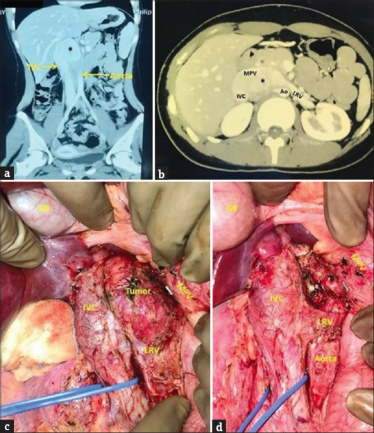
Figure 1.

(a and b) Contrast-enhanced computed tomography abdomen (a) coronal section, (b) axial section showing a well-defined, heterogeneously enhancing mass (asterisk) in the right paravertebral region (D12-L2), closely abutting and displacing the main portal vein (MPV) anteriorly, inferior vena cava (IVC) laterally, left renal vein (LRV) inferiorly and aorta (Ao) posteriorly (c) intraoperative photograph showing tumor enveloped by IVC anterolaterally, MPV anteromedially, Ao posteriorly and LRV inferiorly (d) tumor bed after complete excision showing multiple small arterial and venous branches to the tumor from IVC, Ao, MPV which have been ligated with silk sutures and divided; GB: Gall bladder. MPV: Main portal vein, IVC: Inferior vena cava, LRV: Left renal vein, Ao: Aorta
