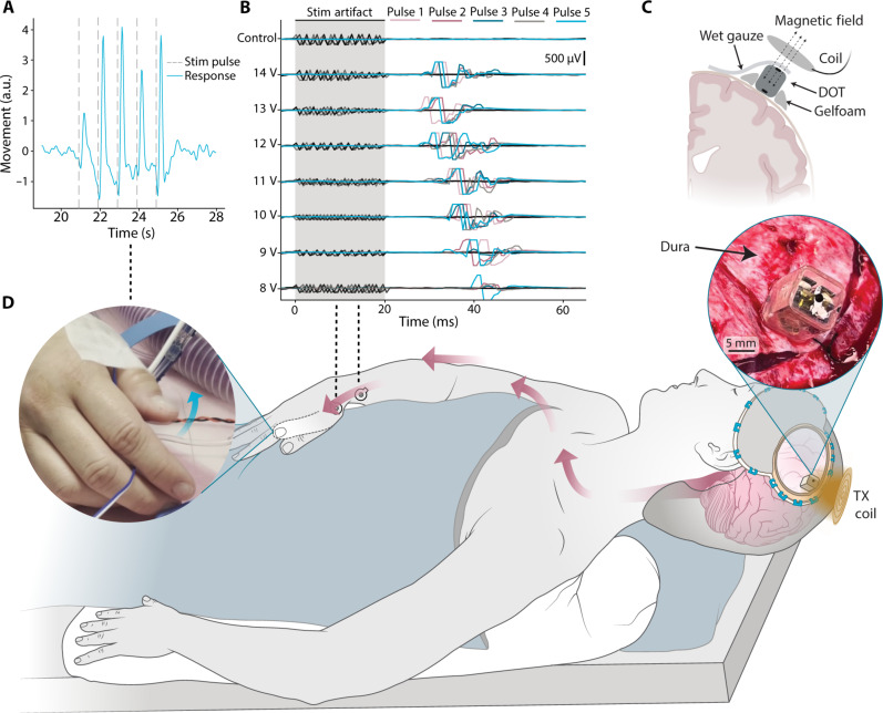
Fig. 4. Intraoperative demonstration of epidural cortical stimulation with a millimeter-sized battery-free device.
(A) Displacement of the thumb extracted from videos during epidural cortical stimulation shows movement in response to each stimulation pulse train applied by the DOT (vertical dashed lines). (B) EMG traces recorded from 5 pulses each in the APB-ADM muscle groups during stimulation at varying amplitudes. (C) Schematic of the intraoperative placement of the device. Saline wetted gauze is used to make electrical connection with the top electrode. (D) A schematic of the intraoperative human studies, where we placed the device above the dura on the motor cortex and activated it with the transmitter coil. Stimulation resulted in contralateral thumb movement. The left inset shows a frame of the movie (movie S2) used to analyze the thumb movement, and the right inset shows a photograph of the DOT placed over the dura. a.u., arbitrary units.

