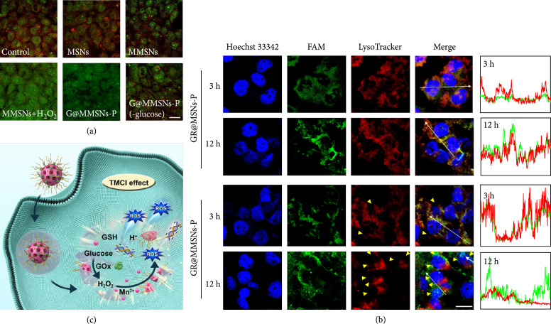
Figure 4.
Endo-/lysosomal disruption capability of GR@MMSNs-P. (a) CLSM images of 4T1 cells after incubation with MSNs, MMSNs, MMSNs+H2O2, G@MMSNs-P, and G@MMSNs-P under low-glucose DMEM medium for 12 h, followed by staining with AO. Lysosome membrane permeabilization was determined by the decreased AO red fluorescence. Scale bar, 25 μm. (b) Endo-/lysosomal escape behavior determined by CLSM images of 4T1 cells treated with GR@MSNs-P and GR@MMSNs-P for 3 and 12 h. FAM-labeled siRNA was shown in green. Lysosome was stained with LysoTracker™ Deep Red. Yellow triangles represented the withered endo-/lysosomes. Scale bar, 20 μm. (c) Schematic illustration of the endo-/lysosomal escape of GR@MMSNs-P.

