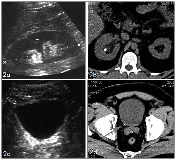
Figure 2. 33-year old man with right-sided renal colic. Noncontrast helical computed tomography (CT) and abdominal ultrasound (US) in the same patient. (a) Coronal identification of the right kidney (between calipers) with a renal stone and mild intrarenal collecting system dilatation. The same findings were found on CT scan (b). In the US scan of the bladder and right ureterovesical junction (c), the radiology resident did not find the small stone (3 mm), shown in CT (d).