
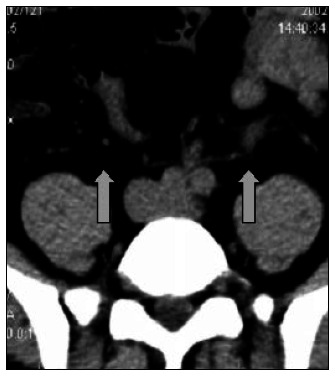
Figure 4. Collecting system dilatation. Noncontrast helical computed tomography (NCT) on a patient with a stone in the distal left ureter, showing ureteral dilatation in comparison with the contralateral normal side (arrows).
Official websites use .gov
A
.gov website belongs to an official
government organization in the United States.
Secure .gov websites use HTTPS
A lock (
) or https:// means you've safely
connected to the .gov website. Share sensitive
information only on official, secure websites.