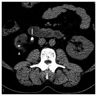
Figure 5. Ureteral fat stranding. Axial slice in noncontrast helical computed tomography (NCT). Stone identified in the proximal right ureter (arrow), associated with ureteral wall edema (tissue rim sign) and perinephric fat stranding. A stone in the kidney is also identified (arrow head).