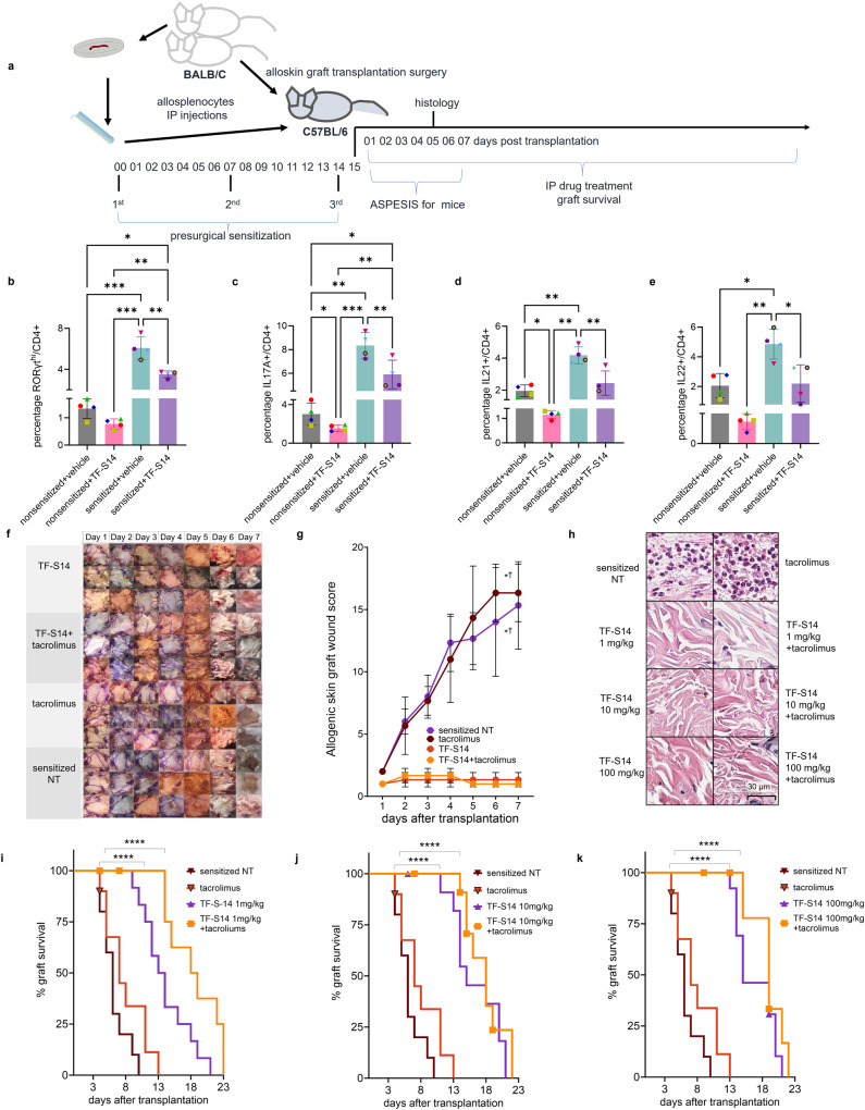
Fig. 3. Effect of RORγt inverse agonist TF-S14 on sensitized mice T-cell to Th17 polarization, allograft survival and histological features of full thickness BALB/C skin transplantation to BALB/C-sensitized C57BL/6 mice.
C57BL/6 were sensitized by three IP injections of BALB/C splenocytes (107) on day 0, 7 and 14. On day 15 mice were transplanted with 12 × 12 mm BALB/C dorsal skin grafts (a). The sensitization was assessed by polarizing T cells isolated from splenocytes of non-sensitized and sensitized mice to Th17 phenotype for 4 days in the presence or absence of TF-S14 (15 nM), followed by stimulation with PMA/ionomycin/monensin/prefoldin cocktail for 5 h. The percentages of RORγthi/CD4+ (b), IL17A+/CD4+ (c), IL21+/CD4+ (d) and IL22+ (e) were measured by flow cytometry. Data are presented as mean ± SD, *p < 0.05, **p < 0.01, ***p < 0.005, REML and Tukey’s tests, n = 4 per group. Following skin BALB/C allograft surgery, pre-sensitized C57BL/6 mice were assigned into sensitized no treatment (NT), tacrolimus (0.5 mg/kg), TF-S14 (1mg/kg) or TF-S14 (1mg/kg) and tacrolimus (0.5 mg/kg) combination daily treatment groups. The allografts were inspected daily, and area (12 × 12 mm) were imaged with 12 megapixels camera (f). The graft area was scored for swelling (1–5), redness (1–5), presence of serous exudate (1–5), presence of purulent exudate (2–10) and loss of hair (2–10) to determine ASPESIS wound score for mice over time. Data are presented as mean ± SD, *†p < 0.05 compared to sensitized NT and tacrolimus treated groups, respectively, Friedman one-way ANOVA and Dunn’s test, n = 3 per group (g). Histopathology photomicrographs of skin allografts isolated on day 5 post-transplantation (H&E, ×40) show that grafts from mice treated with TF-S14 1, 10 or 100 mg/kg alone or in combination with tacrolimus 0.5 mg/kg have low neutrophilic infiltration in <10% of graft area compared to high neutrophilic infiltration (>10%) in sensitized NT mice (h), p < 0.05, Kruskal–Wallis one-way ANOVA and Dunn’s test, n = 3–4 per group. Graft survival assessed daily until complete necrosis or graft loss (i–k). Data are presented in Kaplan–Meier survival curve, ****p < 0.001, Log-rank (Mantel–Cox) test, n = 8–12 per group.

