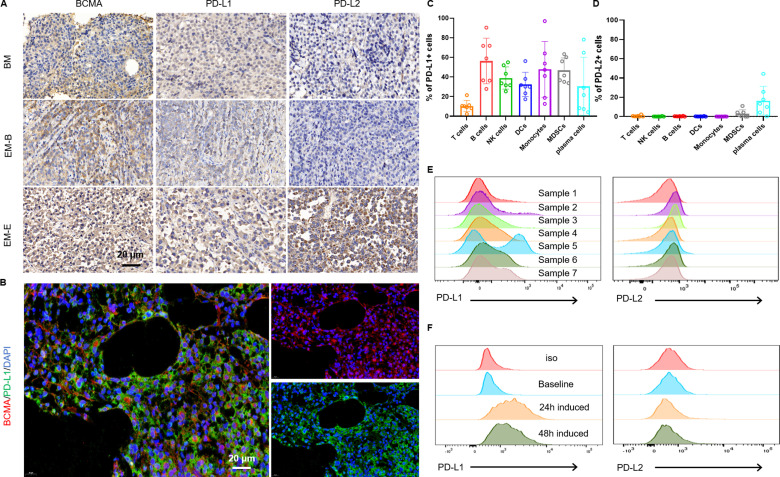
Figure 1.
Characterization of BCMA/PD-L1/PD-L2 expressions in the bone marrow and extramedullary disease sites of MM patients. (A) IHC staining of BCMA/PD-L1/PD-L2 expressions in patient tumor tissues. Representative images of extramedullary bone-related disease arising from the hand and extramedullary extraosseous disease arising from intracranial plasmacytoma. (B) Representative immunofluorescence images showing the expression of BCMA and PD-L1 and their colocalization in BM samples from RRMM patients. Scale bar, 20 µm. Flow cytometry assays showing expressions of PD-L1(C) and PD-L2 (D) on various cell types in BMMCs from seven RRMM patients. (E) Representative histograms of PD-L1(left) and PD-L2 (right) expression in BMMC samples from seven RRMM patients. (F) Representative results showing induced PD-L1 expression in BMMCs after 24 hours coculture or 48 hours of coculture with BCMA CAR-T cells (n=3 independent healthy donors). BCMA, B cell maturation antigen; EM-B, extramedullary bone-related; EM-E, extramedullary extraosseous; IHC, immunohistochemical; MM, multiple myeloma; RRMM, relapsed/refractory MM.

