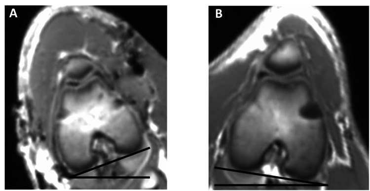
Figure 5. Rotational difference of the distal femur on MRI: (A) left operated femur and (B) right non-operated femur.
MRI images of the distal femora in the operated and non-operated femur showing the difference in the femoral version. For illustrational purposes, both femoral necks are aligned parallel to the axis of the table revealing the difference in the distal femur.

