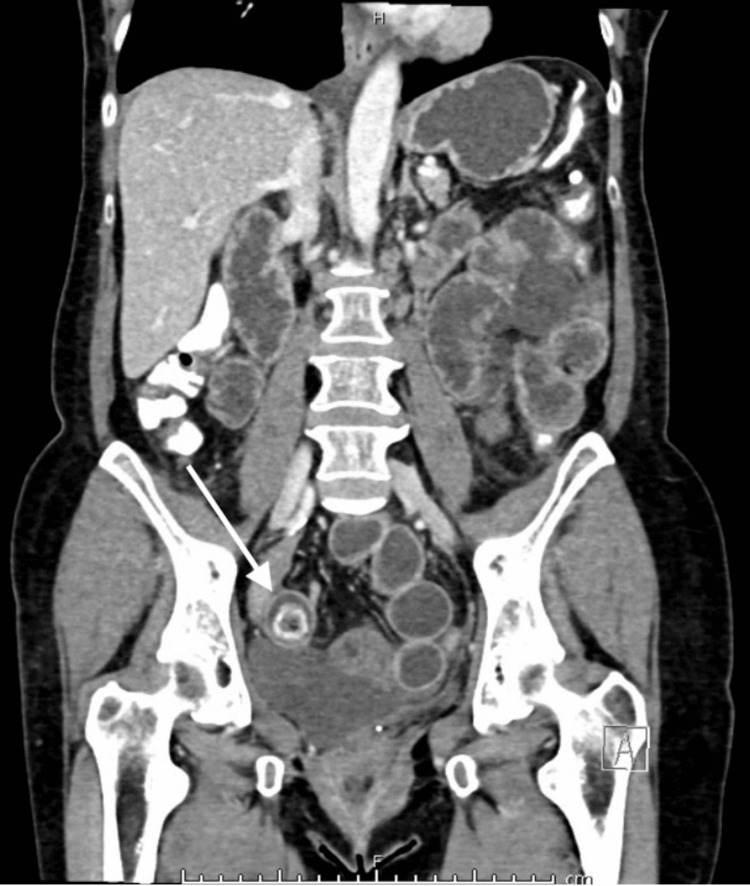
Figure 3. Repeat CT AP during the patient’s second admission.
CT AP: Computed tomography scan of the abdomen and pelvis
This coronal view of CT AP demonstrates high-grade small bowel obstruction secondary to an enterolith (white arrow), which has migrated to the distal ileum from a jejunal diverticulum.

