
Fig. 1.
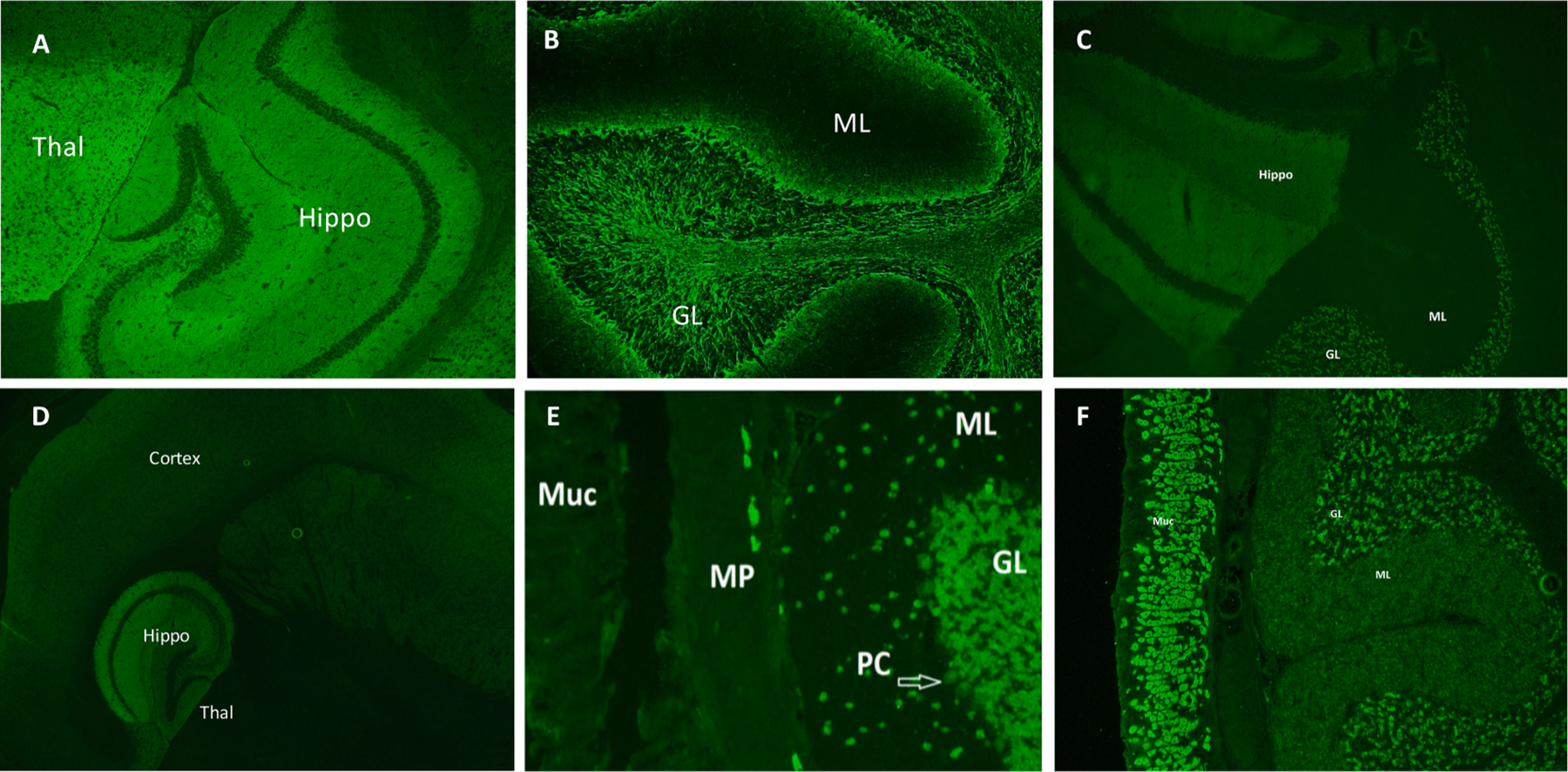
Tissue-based indirect immunofluorescence assay staining features of selected neural antibodies. Synaptic staining of the hippocampus and thalamus (image A, shown) alongside staining of the molecular layer of the cerebellum is characteristic of GABA-B receptor IgG. Neuronal intermediate filament (NIF) IgG demonstrates a classic ‘arborization’ pattern in the granular layer of the cerebellum with a dark molecular layer (image B, shown). NMDA-R IgG stains the hippocampus and the granular layer of the cerebellum (image C). A striking characteristic of AMPA-R IgG is the intense staining of the hippocampus compared to the adjacent thalamus (image D). ANNA-1 IgG demonstrates a nuclear staining pattern with staining of the nucleus and cytoplasm, with sparing of the nucleolus. The nerves of the myenteric plexus also stain which is its sole differing staining feature from ANNA-2 IgG (image E). GAD-65 IgG is visible on TIFA at high titers with punctate staining of the granular layer of the cerebellum (shown), with prominent globus pallida pars interna staining (not shown). It frequently co-exists with gastric parietal cell (GPC) IgG which stains the gastrointestinal mucosa (shown, image F) Thal: thalamus; Hippo: hippocampus; ML: molecular layer; GL: granular layer; Muc: mucosa; MP: myenteric plexus; PC: Purkinje cells.