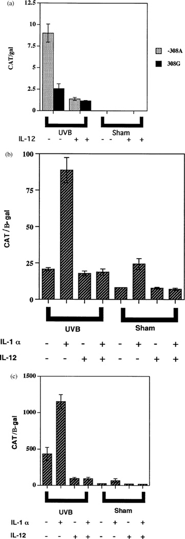
Figure 5. IL-12 inhibits the −308A and −308G TNFα promoter.
Relative activities of TNFα−308G/β-gal construct following transfection into cells. Cells were irradiated with 30 mJ per cm2 of UVB or sham irradiated, and then given media without (minus symbols) or with (plus symbols) 10 ng IL-12 per ml, 10 ng IL-1α per ml (for 3T3 cells only), followed by 24 h of incubation at 37°C. Displayed are normalized levels of CAT expressed by the TNFα promoter construct. The bars represent means (±SEM) of triplicate transfections. (a) Human adult keratinocytes transfected with −308A and −308G TNFα promoter. (b) 3T3 cells transfected with −308G TNFα promoter. (c) 3T3 cells transfected with −308A TNFα promoter.

