
Figure 1.
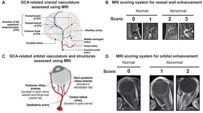
Scoring of cranial and orbital enhancement for GCA. (A) Illustration of vascular anatomy included in MRI scoring for GCA. Arteries included in analysis were selected based on prior knowledge of GCA, including data from postmortem studies. (B) Semiquantitative 4‐point scoring system was used to grade vessel wall contrast enhancement. Images demonstrate examples of each score, ranging from 0 to 3 (2–3 is abnormal). (C) Illustration of orbital vasculature and structures that were scored. Except for the ophthalmic artery, each artery is shown with its corresponding location in parentheses. Additional structures, such as the lacrimal gland and the optic chiasm, were also assessed but are not shown in the figure. (D) Semiquantitative 3‐point scoring system was used to grade contrast enhancement of orbital structures. Images demonstrate examples of each score using optic nerve sheath as an example. Scores range from 0 to 2 (1–2 is abnormal). Grading of the ophthalmic artery wall was according to the same 0 to 3 scoring system used for cranial arteries. GCA, giant cell arteritis; MRI, magnetic resonance imaging; STA, superficial temporal artery.