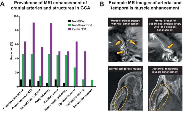
Figure 4.

Multiple arteries and structures with MRI enhancement in participants with GCA. (A) Bar graph of the proportion of patients with MRI enhancement of each artery or muscle in patients without GCA (non‐GCA), with non‐ocular GCA, and with ocular GCA. For each individual arterial segment or structure, there was a higher proportion with MRI enhancement in the GCA versus non‐GCA groups (all P < 0.05). When comparing non‐ocular GCA to ocular GCA, the only structure with a significant difference between groups was the frontal branch of the STA (P = 0.03). (B) Example MRIs demonstrating long‐segment vessel wall enhancement of multiple cranial artery branches (arrows) as well as normal versus abnormal temporalis muscle enhancement (circle), which was observed in 32% of patients with GCA. GCA, giant cell arteritis; MRI, magnetic resonance imaging; STA, superficial temporal artery.
