Figure 5.

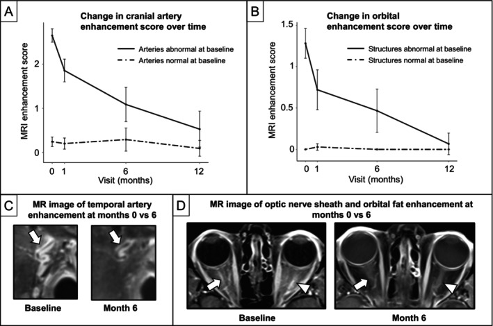
MRI enhancement score decreases over time with treatment of GCA. (A and B) Line graphs with 95% confidence intervals of mean MRI enhancement scores over time separated by baseline normal (dashed line) versus abnormal (solid line) enhancement. We conducted a multilevel mixed‐effect regression analysis for each cranial artery that did not undergo biopsy (branches of superficial temporal artery, occipital artery, and ophthalmic artery) and each orbital structure (optic nerve sheath, intraconal orbital fat, retrobulbar fat at posterior globe, and optic nerve) among 14 patients (11 with GCA, 3 with non‐GCA) who underwent at least one follow‐up MRI after baseline. Both (A) cranial arteries that did not undergo biopsy and (B) orbital structures demonstrated a significant decrease in score over time. Notably, MRI scores were still significantly elevated even after one month of treatment with glucocorticoids (P < 0.01). Example MRIs comparing MRI enhancement at month 0 versus 6 within the same individual for the (C) superficial temporal artery and (D) optic nerve sheath and intraconal fat. GCA, giant cell arteritis; MRI, magnetic resonance imaging.
