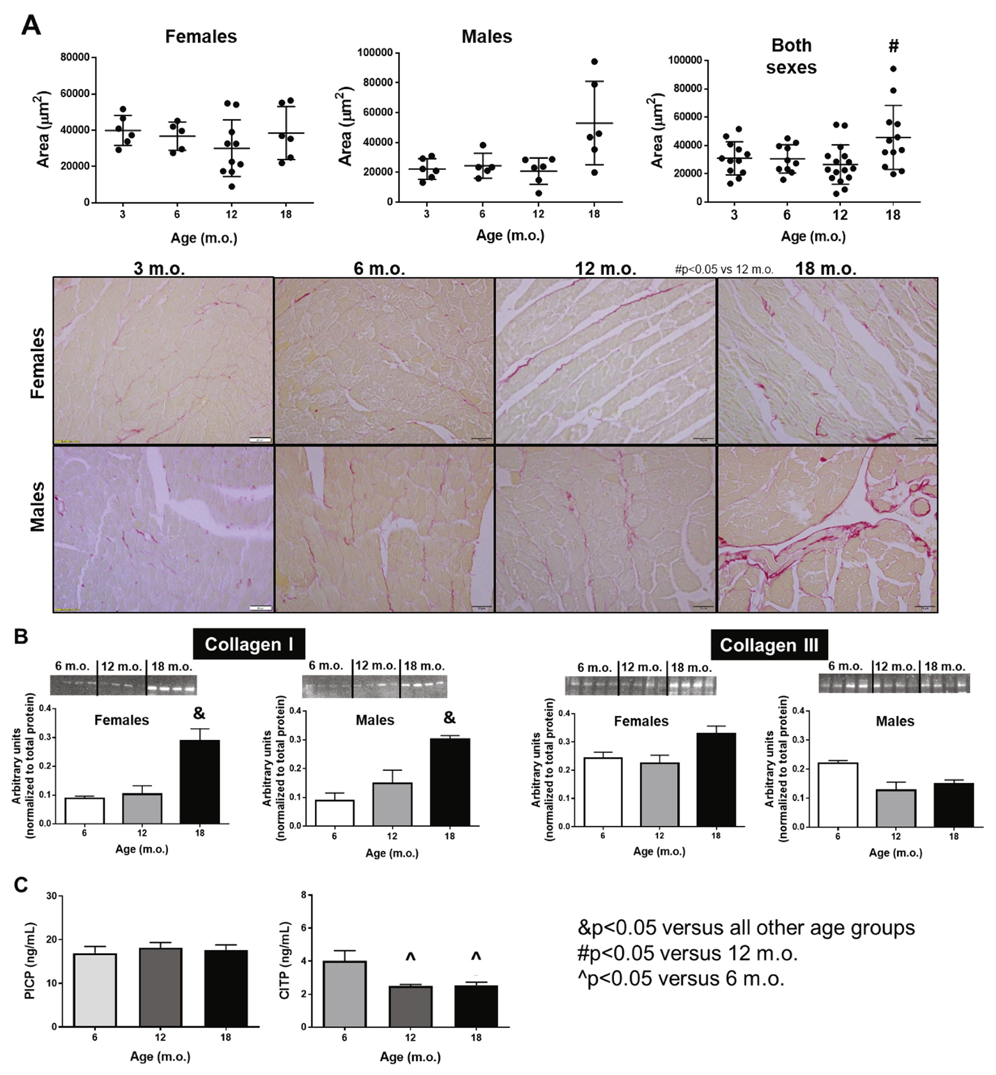
Fig. 5.

Interstitial collagen deposition increases with age in a sex-independent manner. A) Histological quantification of interstitial collagen deposition quantified by picro sirius red staining. B) Collagen type I and III protein expression measured by immunoblot. C) Plasma levels of PICP (collagen synthesis) and CITP (collagen degradation). n = 6/sex/age. (For interpretation of the references to colour in this figure legend, the reader is referred to the web version of this article.)
