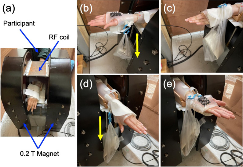
Fig. 1.
The MRI experimental setup. a Top view of the 0.2 T permanent magnet, a1H solenoidal RF coil, and position of participant with his/her forearm during MRI measurement. b, c The cuff setting employed for the supination exercise. The cuff was composed of an opaque, low density polyethylene material, and a weight (5, 15 or 25% of the MVC) was attached on the palmar side of the cuff. The initial position of the hand was set in the fully pronated position (b). The forearm supinated against the weight, and the participants kept the fully supinated position for 1 s (c), then, returned passively to the fully pronated position. This exercise was repeated at 2 s intervals until the participant was unable to continue the forearm rotation. d, e The cuff setting employed for the pronation exercise. A weight was attached on the opisthenar side of the cuff. The initial position of the hand was set in the fully supinated position (d). The forearm pronated against the weight, and the participants kept the fully pronated position for 1 s (e), then, returned passively to the fully supinated position. The yellow arrows show the gravity direction

