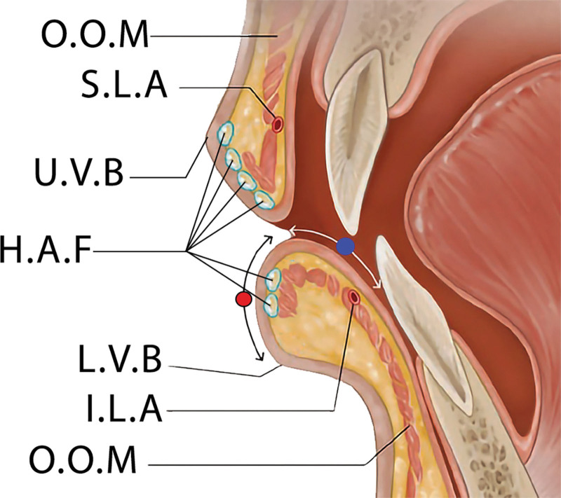
Fig. 2.
Sagittal section illustration of the lip demonstrates the ideal superficial placement for hyaluronic acid fillers. Key anatomical landmarks are labeled to guide the precise administration of the filler. “O.O.M.” denotes the orbicularis oris muscle, which encircles the mouth and is fundamental for lip movements. “S.L.A” indicates the superior labial artery, which supplies blood to the upper lip. “H.A.F” represents the hyaluronic acid fillers, depicted at the preferred superficial injection sites to enhance lip volume and contour. “U.V.B” is marked as the upper vermilion border, the transitional zone between the lip tissue and the normal skin above the lip. “I.L.A” refers to the inferior labial artery, the blood supply to the lower lip. The illustration emphasizes the importance of anatomical knowledge for aesthetic procedures to ensure both efficacy and safety. The red circle indicates the dry vermilion area, and the blue circle indicates the wet vermilion area.

