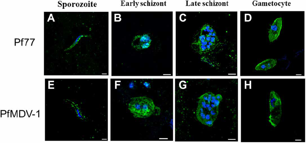
Fig. 4. Pf77 and PfMDV-1 expression in P. falciparum by confocal microscopy.

Expression of Pf77 was assessed in (A) sporozoites, (B) early schizonts, (C) late schizonts, and (D) gametocytes. Expression of PfMDV-1 was also evaluated in (E) sporozoites, (F) early schizonts, (G) late schizonts, and (H) gametocytes. Parasites were stained with mouse anti-Pf77 or mouse anti–PfMDV-1 primary antibody followed by Alexa Fluor 488 AffiniPure Alpaca Anti-Mouse IgG (H+L) secondary antibody and then counterstained with 4′,6-diamidino-2-phenylindole (DAPI). Scale bars, 1 μm.
