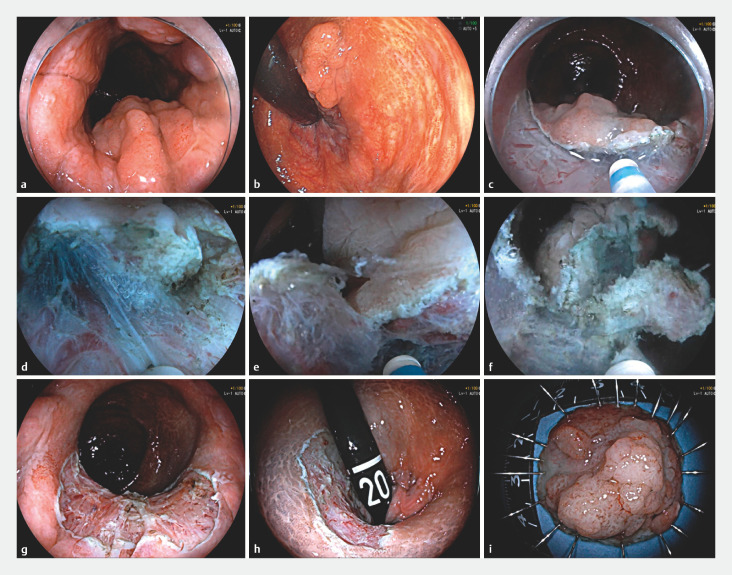
Fig. 2.
a Forward and b retroflexed view of colonoscopy revealed a 25-mm protruding lesion (Paris type 0- Is) in the left wall of the rectum extending close to the dentate line. c The collapsed mucosal flap after a C-shape mucosa incision. d After applying the water pressure method, buoyancy under water immersion provided a countertraction that better exposed the submucosa. The underwater magnified effect also improved visualization during dissection. e The lateral mucosal flap was effectively lifted via active water pressure. f Buoyancy was continuous during the whole procedure. g Forward view showing a minor inner circular muscle injury. h Retroflexed view of the ulcer after resection. i Resected rectal specimen.

