. 2024 Jan 31;14(1):90–107. doi: 10.5455/OVJ.2024.v14.i1.9

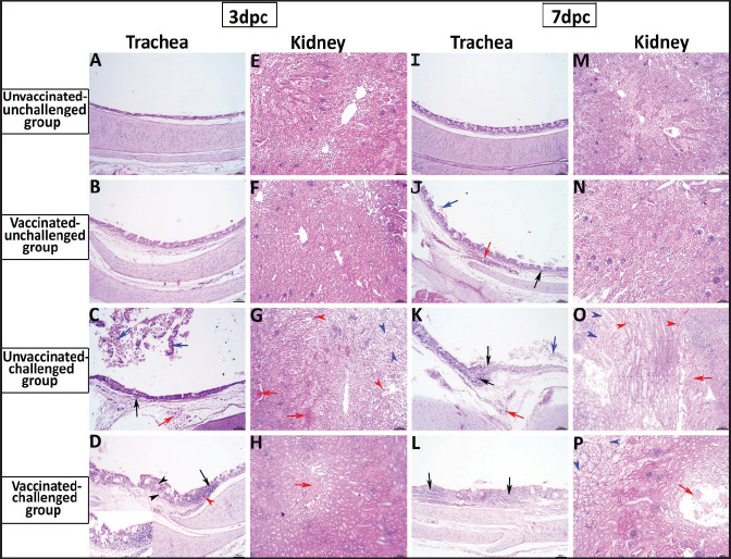
Fig. 5. Photomicrographs of H&E stained tracheal and kidney sections from unvaccinated-unchallenged, vaccinated-unchallenged, unvaccinated-challenged, and vaccinated-challenged chickens at 3 and 7 dpc with variant II IBV strain. (A) Normal tracheal structure. (B) Normal tracheal structure. (C) Trachea showing mucosal congestion (red arrow), inflammatory cell infiltration (black arrow) the presence of luminal necrotic epithelium mixed with mucous and erythrocytes, and desquamation of epithelial lining (blue arrows). (D) Trachea showing hyperplasia of the mucosal glands (black arrowheads), mucosal hemorrhage (red arrowhead), and notable inflammatory cell infiltration (black arrow). (E) Normal structure of the kidney. (F) Normal structure of kidney. (G) Kidney showing vascular congestion (red arrows), interstitial minute hemorrhages (red arrowheads), and vacuolation and necrosis of renal tubules (blue arrowheads). (H) Kidney showing vascular congestion (red arrow). (I) Normal tracheal structure. (J) Trachea showing mild vascular congestion (red arrow), mucosal inflammatory cell infiltration (black arrow), and desquamation of epithelial lining (blue arrow). (K) Trachea showing necrotic lamina epithelia with epithelial desquamation (blue arrow), vascular congestion (red arrow), and notable mucosal infiltration with inflammatory cells (black arrows). (L) Trachea showing notable mucosal inflammatory cell infiltration (black arrows). (M) Normal structure of kidney. (N) Normal structure of kidney. (O) Kidney showing necrotic tubular epithelium (blue arrowheads), vascular congestion (red arrow), and interstitial hemorrhage (red arrowheads). (P) Kidney showing necrotic tubular epithelium (blue arrowheads), and vascular congestion (red arrow)..

Fig. 5.