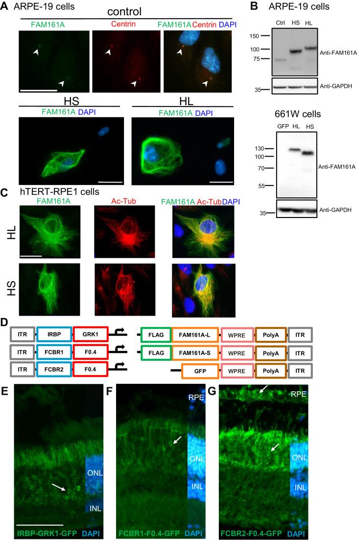
Figure 1. AAV to generate long (L) and short (S) hFAM161A isoforms.
(A) Untransfected ARPE-19 cells (control) were labeled for FAM161A (green) and centrin (red) with arrowheads indicating co-labeled centrioles. To validate both HS and HL FAM161A cDNAs, ARPE-19 cells were transfected with the HL or HS isoforms and labeled for FAM161A (green). The labeling of overexpressed HS and HL was strong and needed a 20-fold decrease in exposure time compared to the labeling of endogenous FAM161A in untransfected cells. (B) Western blot of ARPE-19 and 661 W cells nontransfected (ctrl) or transfected with expression plasmids for GFP, HL, or HS showed the different sizes of the two protein isoforms. (C) hTERT-RPE1 cells were transfected with either the HL or HS isoforms and labeled for FAM161A (green) and acetylated tubulin (red). Overexpression of both isoforms colocalize with acetylated tubulin. (D) Schematic representation of the different elements combined in the AAV8 vectors produced for in vivo studies. Three versions of regulatory elements were followed by either of the three types of transgene elements. (E–G) GFP expression (arrows) one-month post subretinal injection of AAV2/8-IRBP-GRK1-, FCBR1-F0.4-, and FCBR2-F0.4-GFP vectors. Scale bar in (A, C) 25 μm and (E) 100 μm. HS human short isoform, HL human long isoform, Ac-Tub acetylated Tubulin, ONL outer nuclear layer, INL inner nuclear layer, RPE retinal pigment epithelium. Data information: Results in (A) are representatives of four (ARPE-19) and two (661 W) independent experiments. One sample for each condition and each experiment was processed for immunocytochemistry. Data obtained in (C) were generated from one single experiment. Results in (E–G) are representative of 5, 12, and 5 experiments, respectively, with the corresponding eye number processed for IHC: n = 12, 6, and 9. Source data are available online for this figure.

