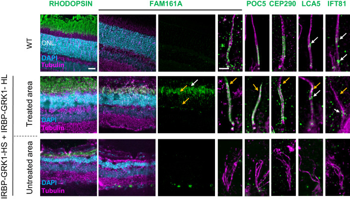
Figure 3. AAV2/8-IRBP-GRK1-FAM161A restores in part the CC structure.
Ultra-tissue expansion microscopy revealed the disorganization of the KO cilium in the untreated area, whereas the co-treatment by the two isoforms restored CC structure organization in the treated area. Note that FAM161A, POC5, and CEP290 localizations are restored in the treated area but their respective labeling extends toward the apical part of the CC (orange arrows) compared to the wild type. In addition, the LCA5 labeling was not grouped, but in part dispersed (orange arrow). IFT81 was also imperfectly distributed (orange arrow) compared to the wild type. For the WT retina, FAM161A labeling from retina cryosection is hardly detectable at low magnification. Scale bar bars: 20 µm and 1 µm for low- and high magnification, respectively. ONL outer nuclear layer, CC connecting cilium. white arrows: correct labeling; orange arrows: ectopic or absence of labeling. Source data are available online for this figure.

