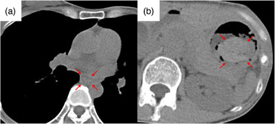
FIGURE 2.

Plain computed tomography scan. (a) A continuous wall thickening from the middle to the lower thoracic esophagus was observed (red arrows). (b) A clearly demarcated hyperabsorptive zone, considered blood in the fundus of the stomach, was observed (red arrows).
