
FIGURE 4.
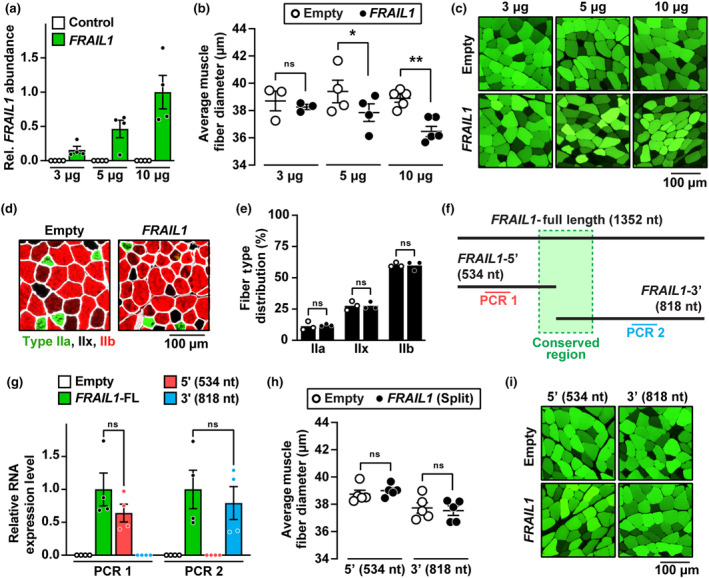
Forced expression of FRAIL1 in mouse skeletal muscle causes muscle atrophy. (a–e) One TA muscle per mouse was transfected with empty plasmid (pcDNA3), and the contralateral TA in each mouse was transfected with plasmid encoding FRAIL1 under the control of the cytomegalovirus (CMV) promoter, as indicated. Seven days posttransfection, bilateral TAs were harvested for RT‐qPCR analysis of FRAIL1 expression (a) histological analysis of skeletal muscle fiber size (b, c) and immunofluorescence microscopy using antibodies targeting MYH2, MYH1, MYH4, and laminin for the quantification of muscle fiber type distributions (d, e). (f) Schematic diagram of the FRAIL1 transcript and synthetic truncated isoforms. The region conserved between nonhuman primates is highlighted in green. (g–i) One TA muscle per mouse was transfected with 10 μg of empty plasmid, and the contralateral TA in each mouse was transfected with 10 μg of plasmid encoding one of the truncated forms of FRAIL1 under the control of the CMV promoter, as indicated. Seven days posttransfection, bilateral TAs were harvested for RT‐qPCR analysis of RNA expression (g) and histological analysis of skeletal muscle fiber size (h, i). Shades of green (c, i) represent differential muscle fiber plasmid uptake following the transfection procedure. Horizontal bars indicate mean values from each group ±SEM, and p values were determined by paired (b, e, h) and unpaired (g) two‐tailed t tests. *p < 0.05, **p < 0.01.