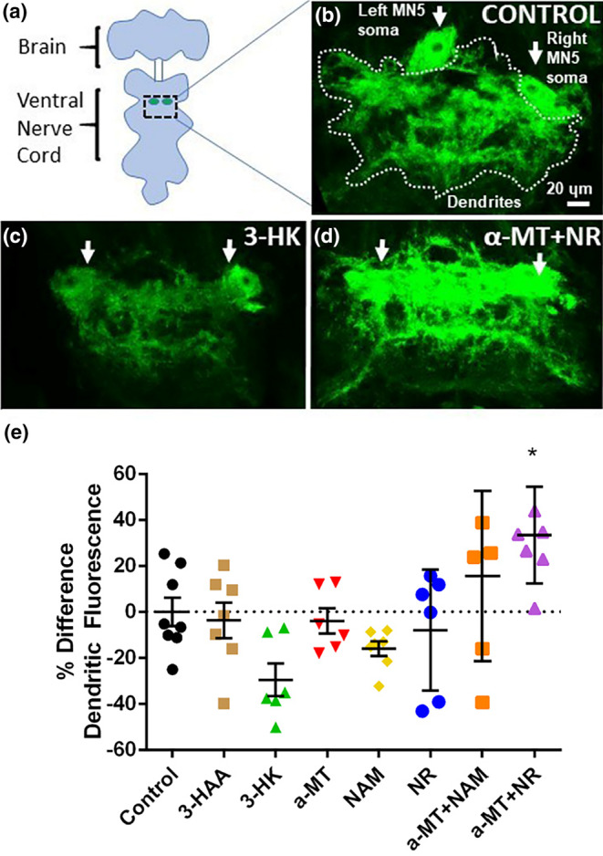
FIGURE 5.

Treatment with kynurenines affects changes in dendritic anatomy. (a) Schematic showing the brain and ventral nerve cord of an adult fly as well as the location of motoneurons that innervate the DLM muscle. Green circles represent cell bodies of the identified motoneuron MN5. Dotted square indicates area of pictures shown in b–d and h–k. (b–d) Projection view images from confocal stacks showing the left and right cell bodies of motoneuron MN5 and the area comprising dendrites from all 10 DLM motoneurons (dotted line) at baseline in flies treated with (b) control food or (c) food supplemented with 3‐HK or (d) α‐MT + NR. (e) Quantification of dendritic fluorescence as calculated by the percentage change of baseline fluorescent value in flies after treatment (n = 6–8 per treatment group). Error bars represent SEM.
