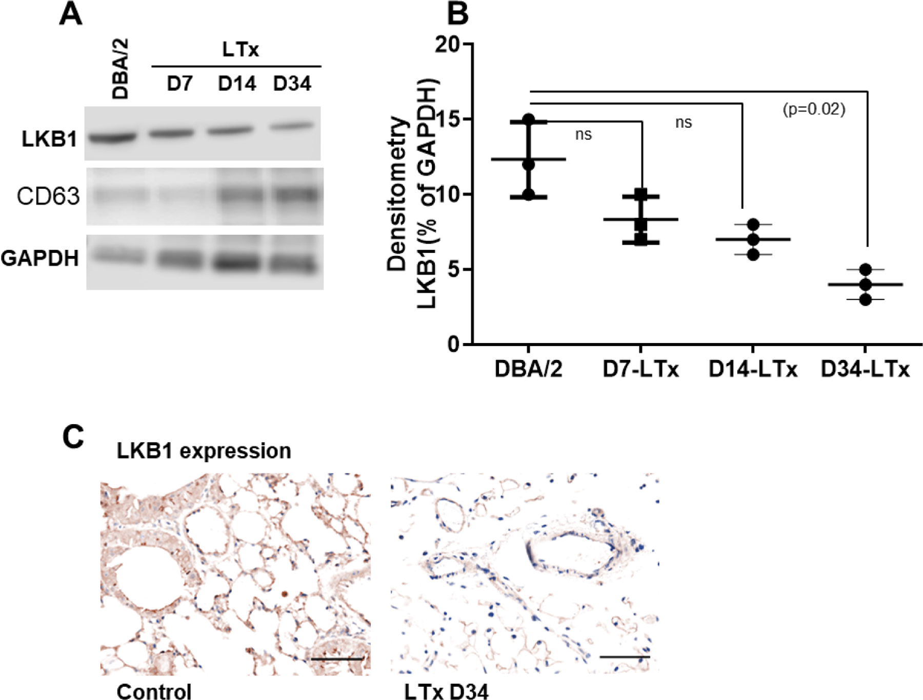
Figure 2:

LKB1 expression in mouse exosome and donor lung tissue. (A) Western blot analysis of LKB1 in exosomal protein isolated from transplanted (day-7, -14, and -34). Control exosome was isolated from non-transplanted DBA/2 mice. CD63 was used as exosome marker, and GAPDH was used as internal loading control. (B) Densitometry analysis showed significant downregulation of LKB1 expression in exosomal protein derived from day-7, -14, and -34 (n=3, p=0.02) after LTx compared to non-transplanted DBA/2 mice. (C) Representative Immunohistochemistry picture of LKB1 expression in donor and recipient lung tissue at day 34 after transplantation. The scale bar is 40x magnification and dimension is 50μm.
