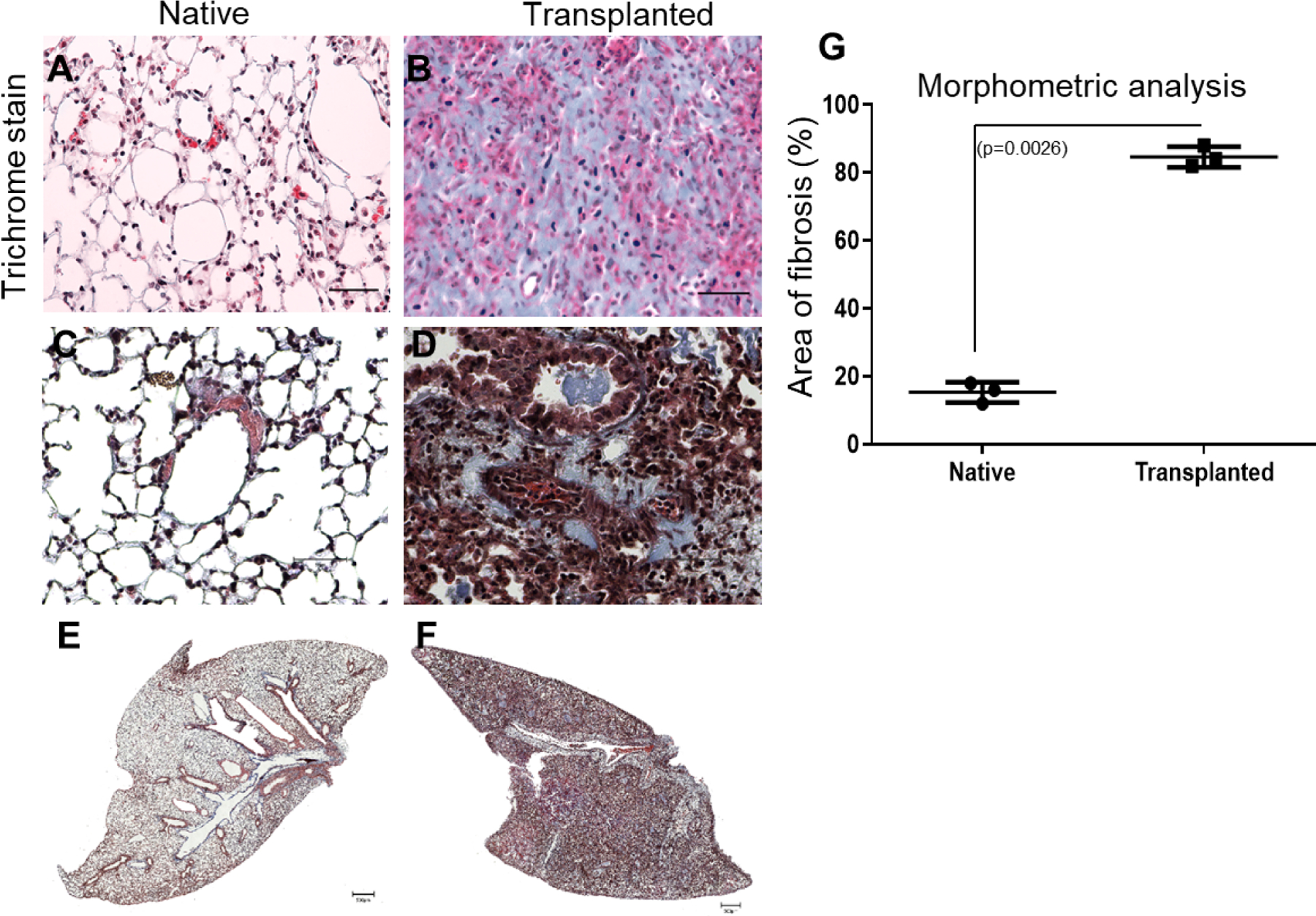
Figure 4:

Histological lesions consistent with chronic rejection and the development of fibrosis in a B6D2F1 to DBA/2 orthotopic LTx murine model as detected by immunohistochemistry. Cut sections were stained with Masson’s trichrome to determine the presence of fibrosis. Masson’s trichrome (A, B) staining in native and transplanted lung tissue. Morphometric analysis of fibrosis: Areas of interest were manually demarcated and measured as a fraction of the total tissue area using ImageJ software (C). Histopathological studies with native lung with normal bronchioles and (D) after murine lung transplantation with perivascular and peri-bronchial fibrosis along with fibrotic plugs. (The scale bar is 40x magnification and dimension is 50μm). Representative Whole lobe scanned image of masson trichrome staining using Invitrogen EVOS M7000 for (E) Native lung (F) Transplanted lung. (The scale bar is 500μm) (n=3).
